Methods
The VRC has expertise in all relevant ophthalmological imaging methods. Our staff members undergo regular training from device manufacturers and are familiar with clinical day-to-day processes due to ongoing contact with clinical staff. This extensive expertise is reflected in our site manuals and our comprehensive investigator support.
OCT (optical coherence tomography)
Optical coherence tomography is a non-invasive imaging technique for acquiring 3D cross sectional scans of the retina. Introduced in the early 1990s, OCT technology has steadily improved over the last decades to become widely accepted with increasing impact in the field of clinical diagnosis and therapy of eye diseases. During an OCT examination the instrument emits a light beam of a specific wavelength into the eye of the patient. The beam is reflected by the different layers of the neurosensory retina, the retinal pigment epithelium (RPE) and choroidal layers. A computer connected to the OCT console calculates the images in respect to the different layers of the retina. An OCT scan displays pathologic characteristics including drusen, intraretinal fluids, cysts, loss of pigment epithelium (geographic atrophy) and numerous other morphological biomarkers which are highly associated with leading retinal diseases such as age-related macular degeneration (AMD), diabetic retinopathies and vascular occlusive disease.
OCTA (optical coherence tomography angiography)
Optical coherence tomography angiography is a relatively new non-invasive imaging technique. Beside the conventional OCT scans that only visualize the static tissues, OCTA also generates 3D scans of the dynamic blood flow in the retinal and choroidal layers. The development of OCTA in the last decade has emerged from the advances in the hardware technology of high-speed spectral domain OCT, that enabled OCTA to take several successive 3D scans within seconds. The differences between the signal intensities of these scans reflect the motion of blood cells. These can be detected efficiently using signal processing algorithms. In this way OCTA is able to identify retinal and choroidal vasculature, and is thus being used clinically to evaluate the progression of diseases such as age related macular degeneration (AMD) and diabetic retinopathy. For instance, due to the detailed 3D view of vasculature provided by OCTA, not only the presence of choroidal neovascularization (CNV) can be analyzed but also its delineation and volumetric size measurements in micrometer-scale.
CF (color fundus photography)
Fundus photography is an imaging modality for capturing photographs of the posterior pole of the eye. A combination of a special microscope and a digital camera provides the necessary platform. The camera can be additionally equipped with a variety of colored image filters. The field of view of the captured area depends on the microscope used and typically ranges from 20 degree up to 60 degree. Mono and stereo images can be acquired with either 3 or 7 fields. The patient’s pupil has to be dilated before an operator can acquire optimal fundus images. The strength of fundus photography lies in the detection of hemorrhages, fibrosis, geographic atrophy, drusen and many more pathologies associated with AMD and diabetic retinopathies.
FA (fluorescein angiography)
For fluorescein angiography, a fluorescent dye is injected into the cubital vein. After the injection, images are taken at defined moments of time to track the distribution of dye in the vasculature of the retina. A flash of light is emitted through a specific image filter into the patient’s eye and excites the fluorescent particles of the dye. The dye begins to distribute within the retinal arteries a few seconds after the injection and in the early arteriovenous phase fills the arterioles and capillary vessels. The dye reaches the retinal veins in the late arteriovenous phase. A healthy eye shows a consistent distribution of the dye through all phases reflected by a normal level of fluorescence of the vessels. The level of fluorescence changes with different pathologies. Hypofluorescence occurs when the flow in the vessels is blocked. Hyperfluorescence occurs when fluorescent particles accumulate in an area, for example through leakage and staining of fluorescein. Fluorescein angiography is an established standard method for detecting a wide range of pathologies that are linked with AMD and diabetic retinopathies.
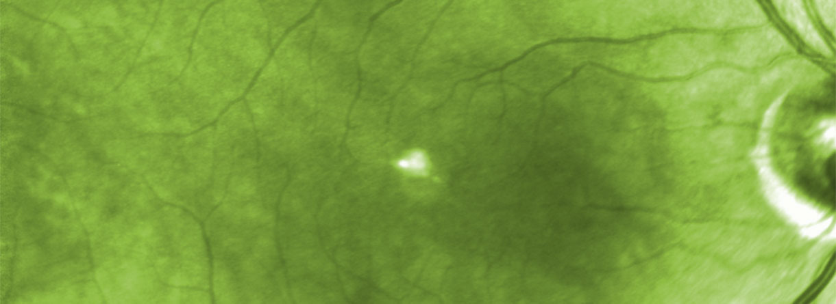
FAF (fundus autofluorescence)
Fundus autofluorescence is a clinical imaging method that can detect early stages of geographic atrophy using the fluorescent properties of specific elements of the retinal layers. It mainly detects lipofuscin, which is a product of the decomposition of photoreceptor cells that aggregates in the retinal pigment epithelium over the years. Lipofuscin produces autofluorescence when excited by light. Areas where it accumulates therefore appear hyperfluorescent on images captured by the fundus camera. In contrast to areas where RPE cells are lost (e.g. in geographic atrophy) present as hypofluorescent.
Indocyanin Green AngiographyCG (ICG)
ICG is a variation of fluorescein angiography. In this examination indocyanin green dye is injected in the patients cubital vein. The dye is than excited with light in the infrared spectrum, and it emits also in this light range that passes through the RPE. This, and the property that in largerly binds to serum proteins (and thus do not extravasate from the fenestrated choroidal vessels) makes it ideal to image the choroidal circulation, and new vessels under the RPE.
MP (microperimetry)
Microperimetry is a special form of computer assisted visual field testing. With microperimetry the retinal sensitivity can be measured, but in contrast to conventional perimetry, here only the central macular retina is tested, and that under constant fundus tracking. This makes a point to point comparison with retinal structures, as well as better follow-up examinations possible.